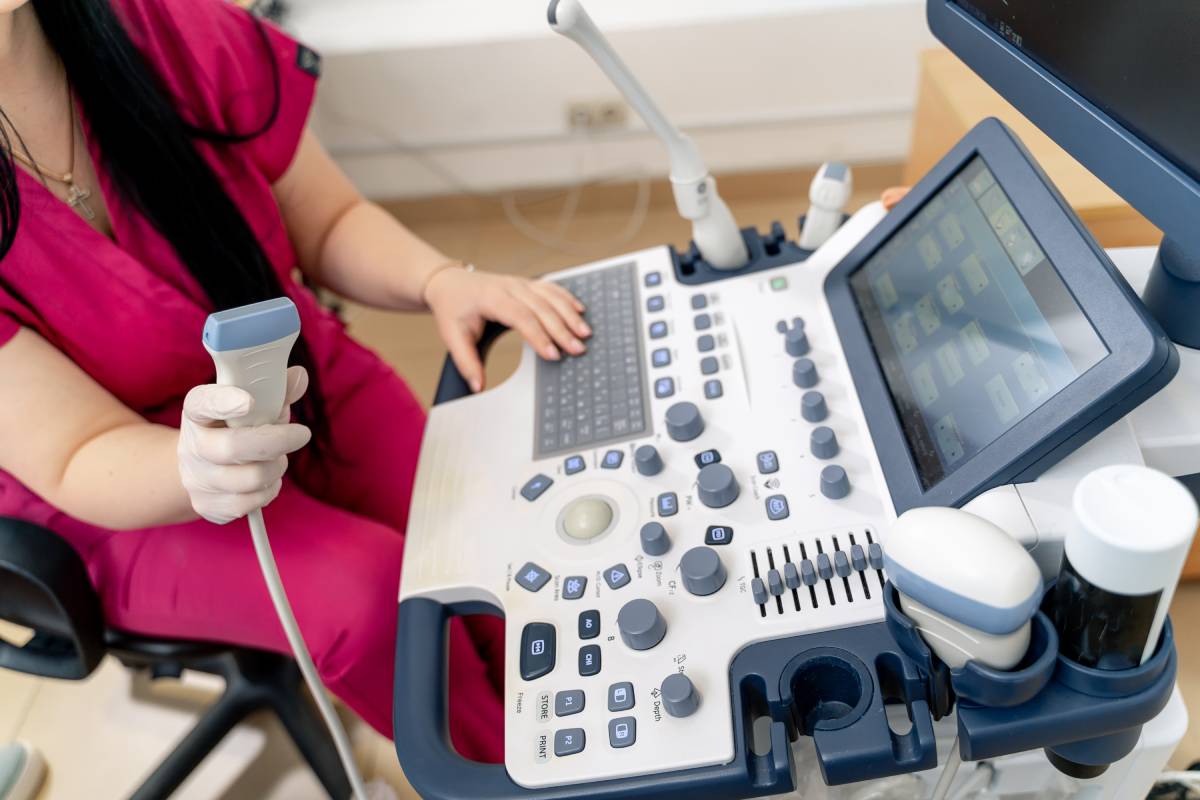
Ultrasound for Spinal Anesthesia in Patients with Obesity

Spinal anesthesia is a type of regional anesthesia that can be used for procedures involving the lower abdomen, pelvic/perineal region and/or lower extremities. Though there are different approaches to administering spinal anesthesia, the general principle involves using anatomy and bony landmarks to safely guide the spinal needle into the ligamentum flavum, dura mater and finally the subarachnoid space without damaging the spinal cord within the meninges. Once in the subarachnoid space, anesthesia is injected into the cerebrospinal fluid resulting in sensory block and subsequent numbness and pain relief [1]. Because accuracy is crucial for spinal anesthesia, ultrasound imaging can help guide needle placement in some situations.
To avoid damaging the spinal cord during this procedure, anatomical knowledge of the spine and surface landmarks are used to approximate the end of the spinal cord. Commonly used landmarks include the vertebral spinous processes and posterior superior iliac crests (PSIS). Identifying these structures can be technically challenging in obese (BMI > 30 kg/m2) or morbidly obese (BMI > 35 kg/m2) patients as they are often impalpable or indistinct [1-4]. An alternative to providing spinal anesthesia is giving general anesthesia. However, general anesthesia requires having adequate airway access, venous access and an understanding of the pharmacokinetic and dosage changes associated with increased adipose tissue in obese patients [5]. Because these factors are more challenging to address in this patient population, general anesthesia is often less appealing than administering regional anesthetics [5-6].
A reasonable solution is using image-guided techniques that enhance the success of safe needle insertion. Ultrasound has been shown to be a safe and effective method for guiding spinal anesthesia, particularly in obese patients [1-4]. One study compared the effect of using bony landmarks to using ultrasound for spinal anesthesia in patients with BMIs of 35 kg/m2 or greater [2]. Their study found that the success rate of inserting the needle the first time using ultrasound was 57%, compared to 30% in patients where only landmarks were used [2]. Multiple other studies have also shown that ultrasound-guided needle insertion reduces the number of needle attempts and increases the success rate of first-needle insertions in patients with difficult to palpate or absent landmarks [1-4] . Generally, in all studies, it was found that ultrasound helps providers by allowing them to determine the correct interspinous spaces, accurately identify the midline and estimate the depth of the dura mater. Lastly, a study by Grau et al., found that using ultrasound was also associated with significant improvements in block-associated pain scores and patient satisfaction.
Ultrasound-guided needle insertion has proven to be advantageous to the traditional landmark approach, however, there are some limitations. Paradoxically, though ultrasound can help elucidate hard-to-palpate structures in obese patients, excess adipose can cause attenuation of ultrasound waves as they travel greater distances through soft tissue to reach bony structures, causing these structures to appear less distinct [2]. One study attempted to circumvent this issue by using fluoroscopic techniques to guide the needle into the subarachnoid space, with success [4]. Using advanced imaging techniques like fluoroscopy can compensate for the imaging limitations of ultrasound, although this procedure is more time consuming, can expose patients and providers to radiation and requires more technical proficiency than using ultrasound [1,4]. Additionally, one study found that in patients with anatomical landmarks that were easy to feel, ultrasound did not improve the first pass needle success rate, and in fact, using ultrasound added unnecessary time to the procedure compared to the landmark approach [7]. Overall, ultrasound has been consistently proven to be a cost and time effective tool for administering spinal anesthesia in obese patients with difficult to palpate spinal anatomy.
References
- Mordecai, M. M., & Brull, S. J. (2005). Spinal anesthesia. Current Opinion in Anesthesiology, 18(5), 527-533. doi: 10.1097/01.aco.0000182556.09809.17
- Chin, K. J., Perlas, A., Chan, V., Brown-Shreves, D., Koshkin, A., & Vaishnav, V. (2011). Ultrasound imaging facilitates spinal anesthesia in adults with difficult surface anatomic landmarks. The Journal of the American Society of Anesthesiologists, 115(1), 94-101.
- Grau, T., Leipold, R. W., Conradi, R., & Martin, E. (2001). Ultrasound control for presumed difficult epidural puncture. Acta Anaesthesiologica Scandinavia, 45(6), 766-771.
- Eidelman, A., Shulman, M. S., & Novak, G. M. (2005). Fluoroscopic imaging for technically difficult spinal anesthesia. Journal of Clinical Anesthesia, 17(1), 69-71.
- Domi, R., & Laho, H. (2012). Anesthetic challenges in the obese patient. Journal of Anesthesia, 26(5), 758-765.
- Pasulka, P. S., Bistrian, B. R., Benotti, P. N., & Blackburn, G. L. (1986). The risks of surgery in obese patients. Annals of Internal Medicine, 104(4), 540-546.
- Jiang, L., Zhang, F., Wei, N., Lv, J., Chen, W., & Dai, Z. (2020). Could preprocedural ultrasound increase the first-pass success rate of neuraxial anesthesia in obstetrics? A systematic review and meta-analysis of randomized controlled trials. Journal of Anesthesia, 34(3), 434-444.
